Chest Radiology > Technique > Positioning > PA VS AP
Positioning
![]()
PA vs AP
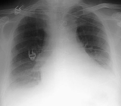
The PA (posterioranterior) radiograph is obtained with the patient facing the cassette and the x-ray tube 6 feet away. This distance diminishes the effect of beam divergence and magnification of structures closer to the x-ray tube. On the image below the exam was obtained in an AP or anteroposterior position. Note that the chest has a difference appearance. The heart shadow is magnified because it is an anterior structure. The pulmonary vasculature is also altered when patients are examined in the supine position. On the AP supine film there is more equalization of the pulmonary vasculature when the size of the lower lobe vessels are compared to the upper.

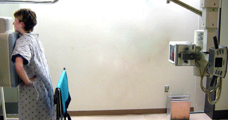
This is the simulated patient in PA (posterioranterior) position.
Note that the x-ray tube is 72 inches away.

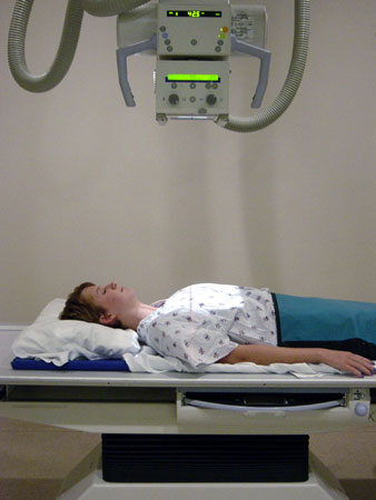
Left, in the supine AP (anteriorposterior) position the x-ray tube is 40 inches from the patient.


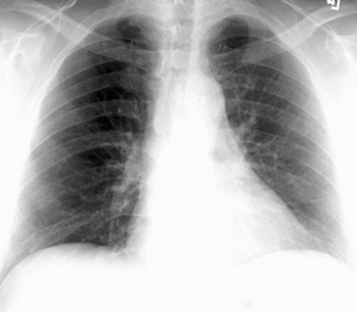
This is a PA upright exam on the left compared with a AP supine exam on the right.
The AP shows magnification of the heart and widening of the mediastinum.
Whenever possible the patient should be imaged in an upright PA position.
AP views are less useful and should be reserved for very ill patients who cannot
stand erect.
