Chest Radiology > Technique > Positioning > Lateral
Positioning
![]()
Lateral Positioning
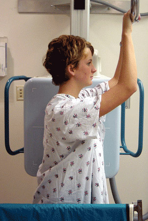
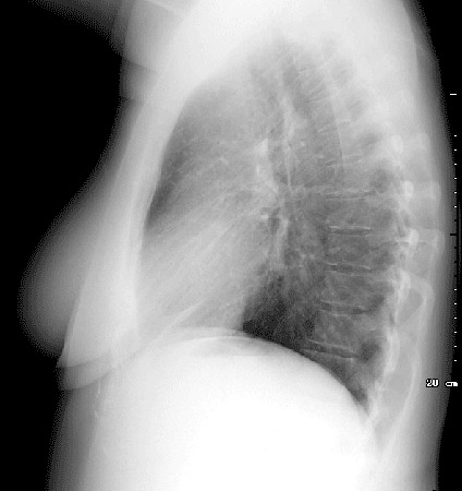
The lateral view is obtained with the left chest against the cassette. This diminishes the effect of magnification on the heart. Looking carefully at the posterior aspect of the chest on the lateral view, which ribs are left and right? Which is the right/left hemidiaphragm?


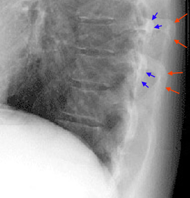
The right ribs (red arrows below) are larger due to magnification and usually projected posterior to the left ribs if the patient was examined in a true lateral position. This can be very helpful if there is a unilateral pleural effusion seen only on the lateral view.

By looking closely at the image you can clearly notice the increased width and posterior location of the right ribs (red arrows) as compared to the left ribs (blue arrows) on CXR.
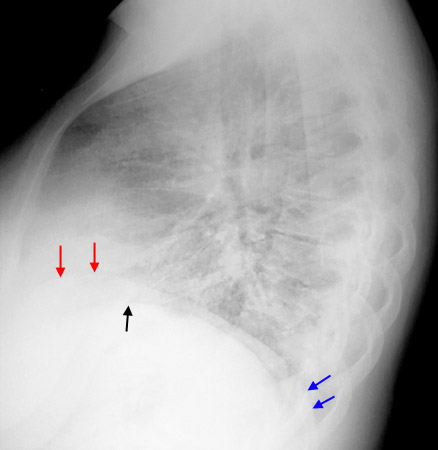
The left hemidiaphragm is usually lower than the right. Also, since the heart lies predominantly on the left hemidiaphragm the result on a lateral radiograph is silouhetting out of the anterior portion of the hemidiaphragm, whereas the anterior right hemidiaphragm remains visible.

Notice how the right diaphragm (red arrows) continues anteriorly, while the left diaphragm disappears (black arrow) because of the silouhetting caused by the heart. Also notice how the right diaphragm at the blue arrows continues past the smaller left ribs and ends at the larger and more posterior right ribs.
