Chest Radiology > Technique > Positioning > PA Lateral
Positioning
![]()
Posterioranterior and Lateral The standard chest
examination consists of a PA (posterioranterior) and lateral chest x-ray. These
images are read together. The PA exam is viewed as if the patient is
standing in front of you with their right side on your left. The patient is
facing towards the left on the lateral view. Comparison images can be
invaluable - Old Gold! If you have comparison exams, the
old PA radiograph is displayed adjacent to the new PA radiograph and the old lateral is
displayed adjacent to the new lateral.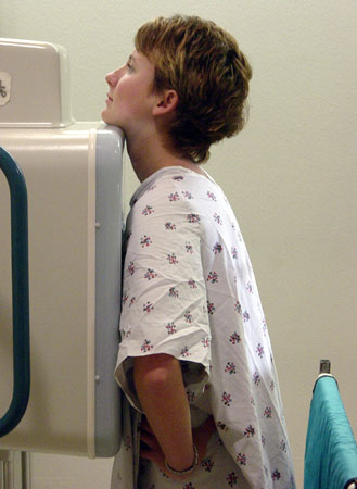
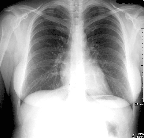
On the left is a simulated patient in position for a standard PA (posterioranterior) chest x-ray. On the right is a normal PA radiograph.
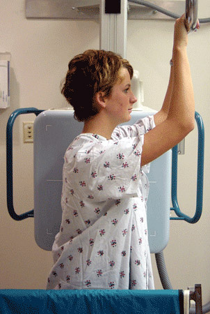
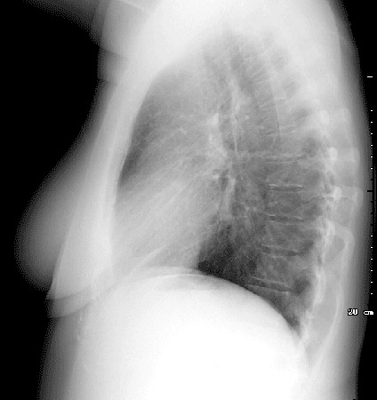
On the left is a simulated patient in position for a lateral chest
x-ray exam and on the right is a normal lateral radiograph.
Note that the receptor or film is against the left chest.


When reading a patient's chest films you should look at both the PA and the lateral rariographs and hang them in this manner (PA on left and lateral on right).
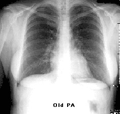
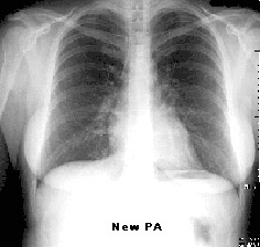
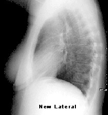
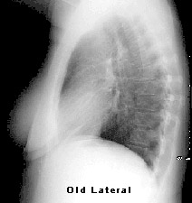
When using comparison exams one should display old PA then new PA then new lateral followed by old lateral. For workstations with 4 monitors, this would be an appropriate display protocol. If your workstation has two monitors, view the PA and lateral, then compare the new PA to old PA, and finally the new lateral to the old lateral.
