Barium Swallow: the hypopharynx & cervical esophagus (cont.)

The Lateral Projection (recorded first)
|
-
Set the digital camera for 4 frames per second.
-
Begin the study with the patient in a lateral position. Aspiration of
barium into the airway is much easier to detect in the lateral view than it
is in the frontal projection.
-
Have the patient take a
mouthful of barium.
-
Center the fluoroscope on the hypopharynx.
Use 9" or 12" FOV. Cone-in side-to-side.
-
Have patient swallow on the count of "three" and watch this first bolus
of barium from the hypopharynx to the GE junction. Quickly rule out
laryngeal penetration or tracheal aspiration by looking at the larynx and trachea. Also,
exclude leakage into the mediastinum or esophageal obstruction.
-
Again, have the patient take a mouthful of barium. Center the fluoroscope
over the hypopharynx and cervical esophagus. (Try to cover an area from the
palate to the shoulders.) Verify that the patient is in a true lateral
position. Have the patient swallow the barium on the count of "three". Begin
recording at 4 frames/sec on the count of "two" and stop when you see on the
monitor that the bolus has passed beyond your field of view.
-
Take one lateral spot image while the patient distends the hypopharynx by phonating "EEEEEE". Zoom to 6" or 9" FOV.
|

|
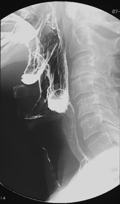
Lateral view of the barium-coated posterior
pharynx and hypopharynx obtained during
phonation demonstrates normal anatomy but
also aspiration of barium into the larynx and
trachea.
|
|
|
|
|
 
|
![]()
![]()