Skeletal Trauma > Introduction > Thoracic and Lumbar Spine Review
Thoracic and Lumbar Spine Review
![]()
|
The spinal trauma covered in this tutorial is limited to that at the thoracic and lumbar levels. Therefore,
this brief review only covers the thoracic and lumbar spine. For complete coverage of the cervical spine, please
refer to the Imaging Evaluation of the Cervical Spine
self tutorial. There are 12 thoracic vertebrae and 5 lumbar vertebrae. Each vertebrae can be divided into several parts. | |
 |
|
|
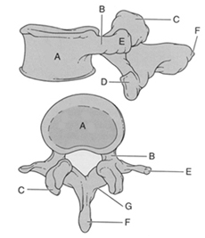
It may be valuable to draw a thoracic vertebra to better understand its anatomy. The drawings below show simple
schematics of superior and lateral views of a thoracic vertebra. | |
 | |
|
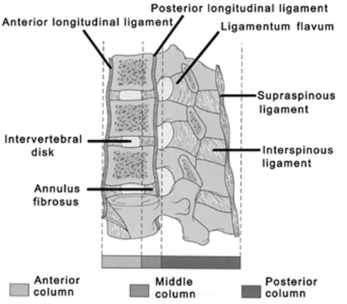
Also note the 4 major ligaments that contribute the the stability of the spine: anterior longitudinal ligament,
posterior longitudinal ligament, ligamentum flavum, and interspinous ligament. | |
 | |
|
One should consider the three columns of the spine for assessment of stability. The anterior column consists
of the anterior longitudinal ligament and the anterior part of the vertebral body. The middle column includes the
posterior vertebral body and the posterior longitudinal ligament. The posterior column consists of the pedicles,
the ligamentum flavum and ther interspinous ligaments. Fractures involving only one column are stable and those
involving all three are unstable. It is important to remember that thoracic and lumbar nerve roots exit from the upper portion of the neural foramen from the vertebral level below (e.g., L4 root exits at the L4-L5 foramen). | |
