The human skeleton contains 206 bones. All of these bones can be classified into five groups based on shape.
Below are the definitions and a few examples of the five bone classifications.
- Long bones - bones of the extremities that have a length greater than the width (e.g., femur).
- Short bones - bones of the wrist, ankle and foot that are cuboidal in shape (e.g., carpals and tarsals).
- Flat bones - diploic bones of the vault of the skull (e.g., parietal and frontal) and the iliac bone.
- Sesamoid bones - small, rounded bones located in tendons (e.g., metatarsophalangeal joint of the great toe).
- Irregular bones - as the name implies, these bones have irregular shapes (e.g., vertebrae, sacrum, coccyx).
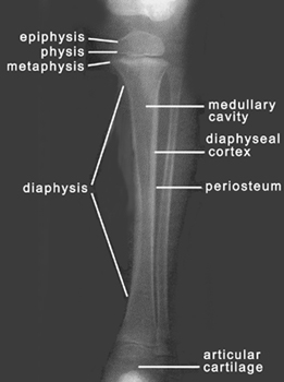
Review the parts of a long bone in a child with the open epiphyses. In an adult, the epiphyses would
either be closed and not seen or evidenced by a sclerotic scar.
|

|
|
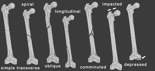
When long bones fracture, there are specific terms for the different patterns.
|

|

|
Here are the definitions for the fracture patterns shown above:
- Simple Transverse Fracture - a fracture in which the fracture line is perpendicular
to the long axis of the bone and that results in two fracture fragments.
- Oblique Fracture - a fracture in which the fracture line is at an oblique angle to
the long axis of the bone.
- Spiral Fracture - a severe form of oblique fracture in which the fracture plane rotates
along the long axis of the bone. These fractures occur secondary to rotational force.
- Longitudinal Fracture - a fracture in which the fracture line runs nearly parallel to the
long axis of the bone. A longitudinal fracture can be considered a long oblique fracture.
- Comminuted Fracture - a fracture that results in more than two fracture fragments.
- Impacted Fracture - a fracture in which the end of the bone is driven into the contiguous
metaphyseal region without displacement. This type of fracture occurs secondary to axial or compressive
force.
- Depressed Fracture - a form of an impacted fracture that involves the articular surface of a bone
and results in joint incongruity.
- Avulsion Fracture - (not pictured above) a fracture in which the tendon is pulled away from the bone,
carrying a bone fragment with it.
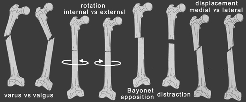
Fractures are also described in terms of alignment. Below are the definitions of the various types of alignment
that result from the fracture patterns just described. The different alignments can also be seen in the above images.
- Varus vs Valgus - varus and valgus deformities are both angulations, which are described according to the
direction of the apex or the direction in which the distal fragment is angled. In varus deformity, there is apex
angulation away from the midline and the distal structure moves medially (i.e., bowleggedness). In valgus deformity,
there is apex angulation toward the midline and the distal structure moves laterally (i.e., knock-kneed).
- Internal vs External Rotation - rotation is described according to the direction of movement of the distal
fragment.
- Bayonet Apposition - overlap of fracture fragments.
- Distraction - longitudinal separation of fracture fragments.
- Medial vs Lateral Displacement - occurs when the cortices are out of alignment. Displacements are described
according to the direction of movement of the distal fragment relative to the proximal fragment.
When describing a fracture, one should describe the location, pattern and alignment. Remember, the alignment is described for the
distal fragment relative to the proximal, with the patient in anatomical position.
|
![]()


