Emergency Ultrasound > RUQ Pain > Gallstones
RUQ Pain - Gallstones
![]()
Clinical
Gallstones affect 10-15% of the population and are a major cause of gallbladder (GB) morbidity. Symptomatic gallstones presents with characteristic right upper quadrant discomfort or pain (biliary colic). Most gallstones are mixtures of cholesterol, calcium bilirubinate, and calcium carbonate.

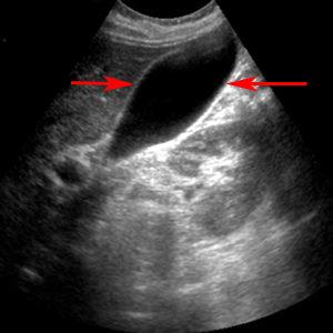
Normal appearing gallbladder indicated between arrows.
Exam
Begin the exam with the patient in the supine position. The patient can be moved to the left posterior oblique or upright position to demonstrate stone mobility. Obtain full length of gallbladder from the portal vein to fundus and transverse images at representative levels. Measure GB wall thickness perpendicular to wall. Obtain full length of common bile duct (CBD) or as much as possible, and measure CBD. Acquire longitudinal and transverse views of pancreatic head. Document any stones or biliary dilatation. If stones are seen, evaluate if they are mobile or impacted. Move the patient into upright or lateral decubitus positions to demonstrate stone mobility.
Sonographic Diagnosis:
1) Echogenic foci in GB lumen
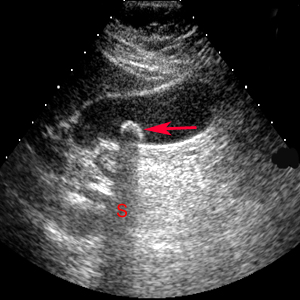
2) Acoustic shadowing

Gallstone (red arrow) within the gallbladder produces a bright surface echo and causes a dark acoustic shadow (S).
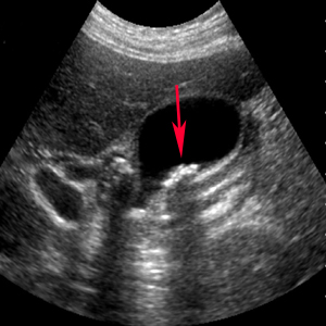
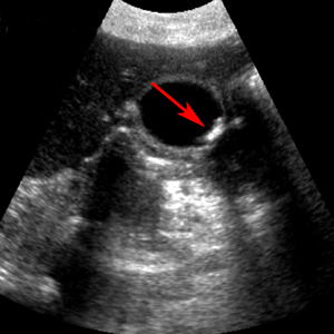
3) Rolling stone sign - movement of gallstones with GB with position change
A B
B
Rolling Stone Sign. A. With the patient supine, the gallstone (red arrow) is near the neck of the gallbladder. B. With the patient in left lateral decubitus position, the gallstone (red arrow) rolls to the gallbladder fundus.
