Emergency Ultrasound > Epigastric Pain > Pancreatitis
Epigastric Pain - Pancreatitis
![]()
Clinical
Acute pancreatitis is most commonly caused by alcohol abuse or a gallstone impacted in the distal common bile duct. Inflammatory changes vary from mild interstitial edema to extensive necrosis with hemorrhage. Patient usually presents with deep epigastric pain that radiates to the back, nausea, vomiting, abdominal tenderness, fever, leuckocytosis, and elevated pancreatic enzymes. Pancreatic pseudocysts are sometimes found several weeks after pancreatitis.

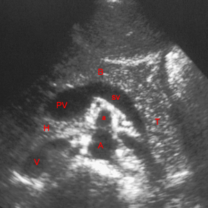
In the transverse image, the pancreas is recognized by identifying its adjacent vasculature: inferior vena cava (V), abdominal aorta (A), and the superior mesenteric artery (a). The junction of the splenic vein (sv) with the superior mesenteric vein marks the commencement of the portal vein (PV) and is recognized by its teardrop shape. The head (H), body (B), and tail (T) of the pancreas course anterior and parallel to the splenic vein (sv).
Exam
Begin with the patient in the supine position or the upright position. Multiple views of the pancreas are required in the transverse plane (long axis of the pancreas) and longitudinal plane with identification of head, uncinate, neck, body and tail. Visualization of the pancreas in patients with a lot of air in the stomach may require additional maneuvers such as filling the stomach with water. In those cases where the pancreas is poorly visualized in spite of additional maneuvers, always document the pancreatic area in both transverse and longitudinal planes.
For a thorough survey of the pancreas, a minimum of six images are required, three transverse and three longitudinal. Depending on the patient, additional pictures may be needed to show the entire pancreas. Documentation of pathology (masses, pseudocysts, nodes) requires additional images.
Sonographic Findings :
1) Diffuse enlargement of pancreas with ill-defined margins and hypoechoic parenchyma
2) Peripancreatic fat decreased in echogenicity with hypoechoic stranding densities
3) Hemorrhage may cause hyperechoic masses of clot of blood
A B
B

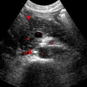
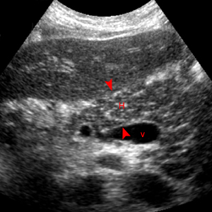
Acute Pancreatitis. A. Transverse scan. B. Longitudinal scan. The head of the pancreas (H) is enlarged as revealed by the red arrowheads and decreased in echogenicity because of edema. The surrounding structures are superior mesenteric vein (v), superior mesenteric artery (a), abdominal aorta (A), and inferior vena cava (IVC).
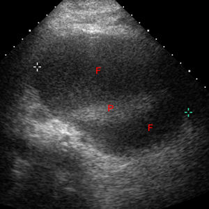
4) Peripancreatic fluid collections in lesser sac, perirenal areas, and small bowel mesentery

Transverse image shows huge fluid collection (F) surrounding the pancreas (P).
