ICU Chest Films > Fluid in the Chest > Pulmonary Edema > Interstitial Edema
Interstitial Edema
![]()
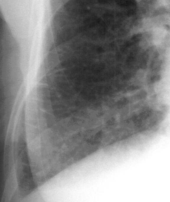
Interstitial edema occurs as venous pressure rises into the 25-30 mmHg range. Interstitial edema as seen on the chest x-ray may in fact preceed clinical symptoms. This is testimony to the importance of the ICU chest film. Alveolar epithelial junctions are much tighter than endothelial cell junctions. Therefore, excess fluid accumulates in the intersitial space surrounding capillary walls first. Several signs are indicative of interstitial edema. The large pulmonary vessels may begin to lose definition and become hazy. Septal lines may begin to appear. Kerley's A lines range from 5 to 10 cm in length and extend from the hila toward the periphery in a straight or slightly curved course. They represent fluid in the deep septa and lymphatics, usually in the upper lobes. Kerley's B lines are shorter thin lines (1.5 to 2.0 cm in length) and are seen in the periphery of the lower lung, extending to the pleura (see below). These represent interlobular septal thickening. The chest x-ray in interstitial edema may take on a diffuse reticular pattern resembling widespread interstitial fibrosis. Peribronchial cuffing represents interstitial edema and appears as very thick bronchial walls.


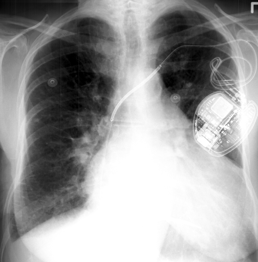
The patient above is suffering from congestive heart failure resulting
in interstitial edema (left). Notice the Kerley's B lines in the closeup
view (right).