Emergency Ultrasound > Technique > Transducer
Technique - Transducer
![]()
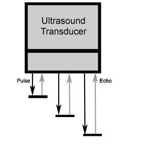
A transducer is a device that translates one form of energy to another. An ultrasound transducer contains a piezoelectric crystal that can translate electrical signals into mechanical energy or mechanical energy into electrical signals. The transducer uses a pulse-echo technique to obtain an image. Initially, a sound wave is produced by electricity within the transducer and directed into the patient. The reflected sound waves are received by the transducer and converted into electrical signals, and an image can be created.

The Ultrasound (US) transducer sends a series of US beams into patient tissue. The US image is produced by the pattern of reflected beams (echoes). The depth of an echo is determined by measuring the round trip time-of-flight from beam transmission to echo reception. Assuming that the speed of sound in human tissue is a constant 1540 m/sec, the depth of an echo can be accurately plotted on the resulting US image.
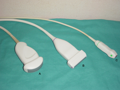
Linear, sector, and curved array are three formats of a transducer that determine the shape and field of view. Linear array transducers produce rectangular images and offer the best overall image quality. Sector array transducers produce slice-of-pie-shaped images and are optimal for examining larger organs from between the ribs. Curved array transducers combine advantages of the sector and linear formats and are optimally used when the sonographic window is large.

Curved (A), linear (B), and sector (C) array transducers provide differing shapes in the ultrasound field-of-view.
Medical ultrasound is performed using very high sound frequencies in the range of 1-20 MHz. The best image resolution is obtained by using the highest transduced frequency possible. However, the higher frequencies are more limited in ability to penetrate tissue. Thus, lower frequencies are often used, accepting lower resolution as a trade-off for better penetration for deeper imaging.
