Chest Radiology > Anatomy > Lobes & Fissures
Lobes
and Fissures
![]()
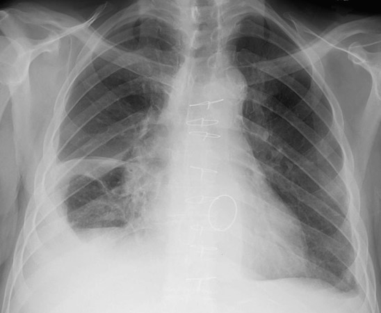
On the PA chest xray, the minor fissure divides the right middle lobe from the right upper lobe and is sometimes not well seen. There is no minor fissure on the left. The major fissures are usually not well seen on the PA view because you are looking through them obliquely. If there is fluid in the fissure, it is occasionally manifested as a density at the lower lateral margin.
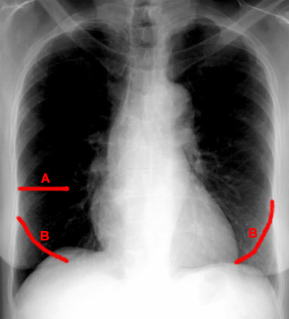
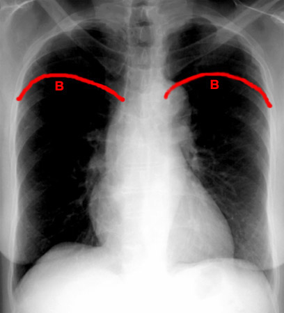
The left image shows the right minor fissure (A) and the inferior borders (B) of the major fissures bilaterally. The right image shows the superior border of the major fissures (B) bilaterally.
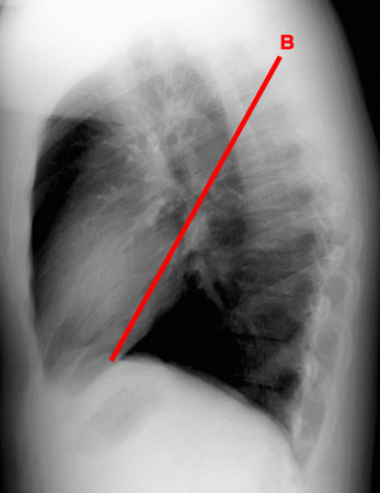
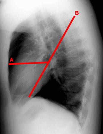
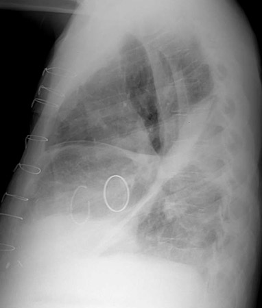
On the lateral view, both lungs are superimposed.
Think about them
separately, the left lung has only a major fissure as shown.
The right lung
will have both the major and minor fissure.
The patient above has a pleural effusion
extending into the fissure.
Which fissure is which?
What is the bright loop near the center of the
films?
( Click each image for answers)
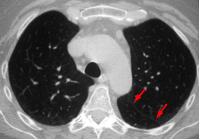
On a CT scan the fissures are shown as an area of paucity of vessels in the region of the capillaries near the fissure. If a very thin slice is taken, the pleura can actually be seen as a line (arrows).

