Imaging of the Cervical Spine > Technique > MRI
MRI
![]()
MRI is indicated in cervical fractures that have spinal canal involvement, clinical neurologic deficits or ligamentous injuries. MRI provides the best visualization of the soft tissues, including ligaments, intervertebral disks, spinal cord, and epidural hematomas.
The advantages of MRI are:
1. excellent soft tissue constrast, making it the study of choice for spinal cord survey, hematoma, and ligamentous injuries.
2. provides good general overview because of its ability to show information in different planes (e.g. sagital, coronal, etc.).
3. ability to demostrate vertebral arteries, which is useful in evaluating fractures involving the course of the vertebral arteries.
4. no ionizing radiation.
The disadvantages of MRI are:
1. loss of bony details.
2. relatively high cost.
At the University of Virginia, the protocol for MRI in cervical spine trauma follows five sequential scans: T1 turbo spin echo in sagittal plane, Turbo T2 in sagittal plane, 2D flash in sagittal plane, 2D flash in axial plane and T1 turbo spine echo in axial plane.
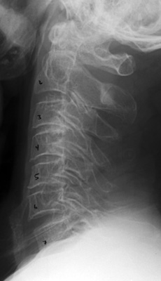
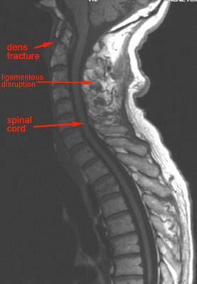
Here is an example of a MRI image of the cervical spine demostrating a ligamentous injury. Notice that the spinal cord is also very well delinated. A dens fracture is not obvious on the lateral film, but is clearly revealed on MRI.


