ICU Chest Films > Lung Processes > Atelectasis > Left Lower Lobe
Left Lower Lobe Atelectasis
![]()
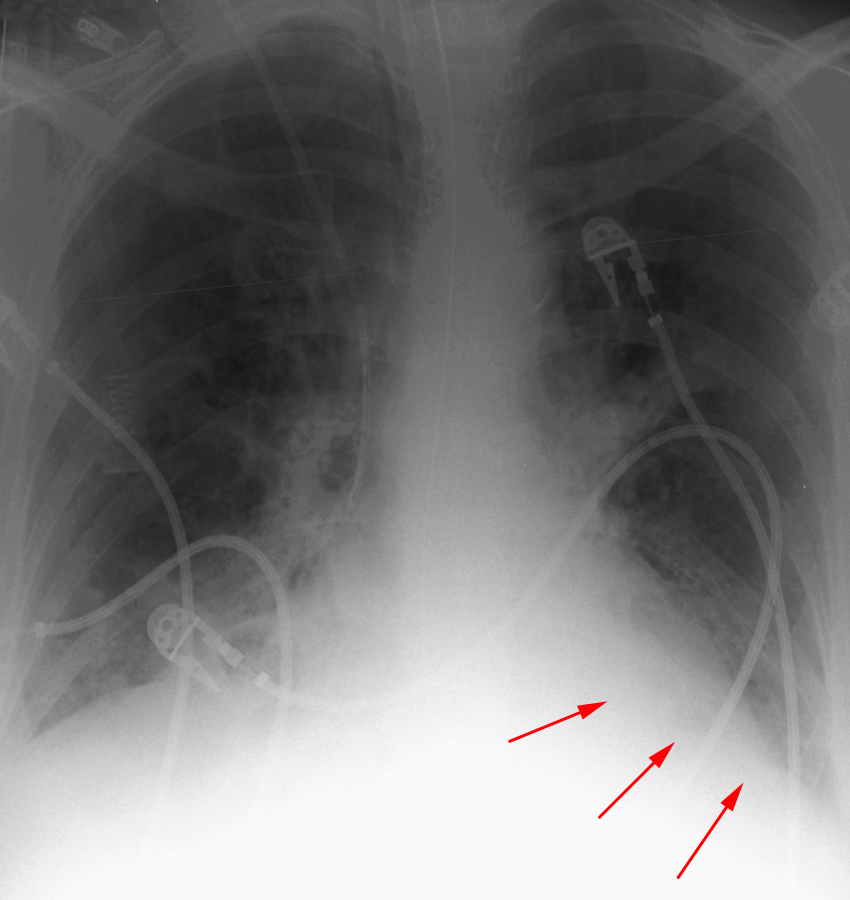
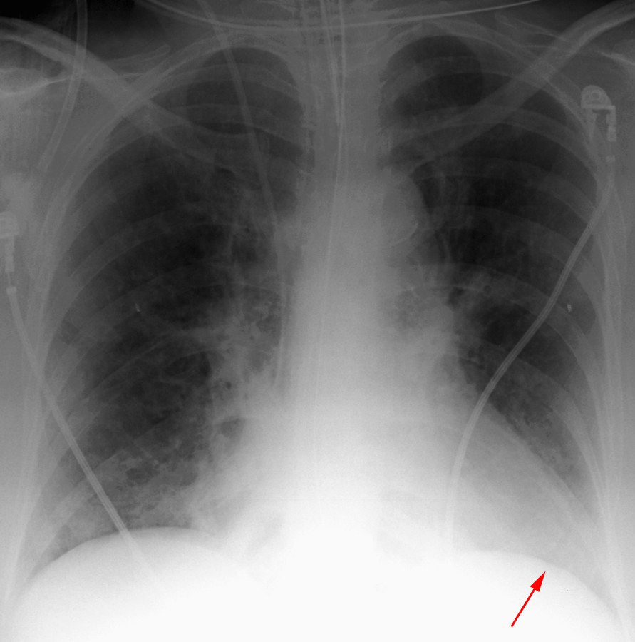
Atelectasis of either the right or left lower lobe presents a similar appearance. Silhouetting of the corresponding hemidiaphragm, crowding of vessels, and air bronchograms are standard, and silhouetting of descending aorta is seen on the left. It is important to remember that these findings are all nonspecific, often occuring in cases of consolidation, as well. A substantially collapsed lower lobe will usually show as a triangular opacity situated posteromedially against the mediastinum.


These radiographs demonstrate left lower lobe atelectasis followed by
resolution, respectively.