ICU Chest Films > Lung Processes > Atelectasis > Radiographic Appearance
Radiographic Appearance of Atelectasis
![]()
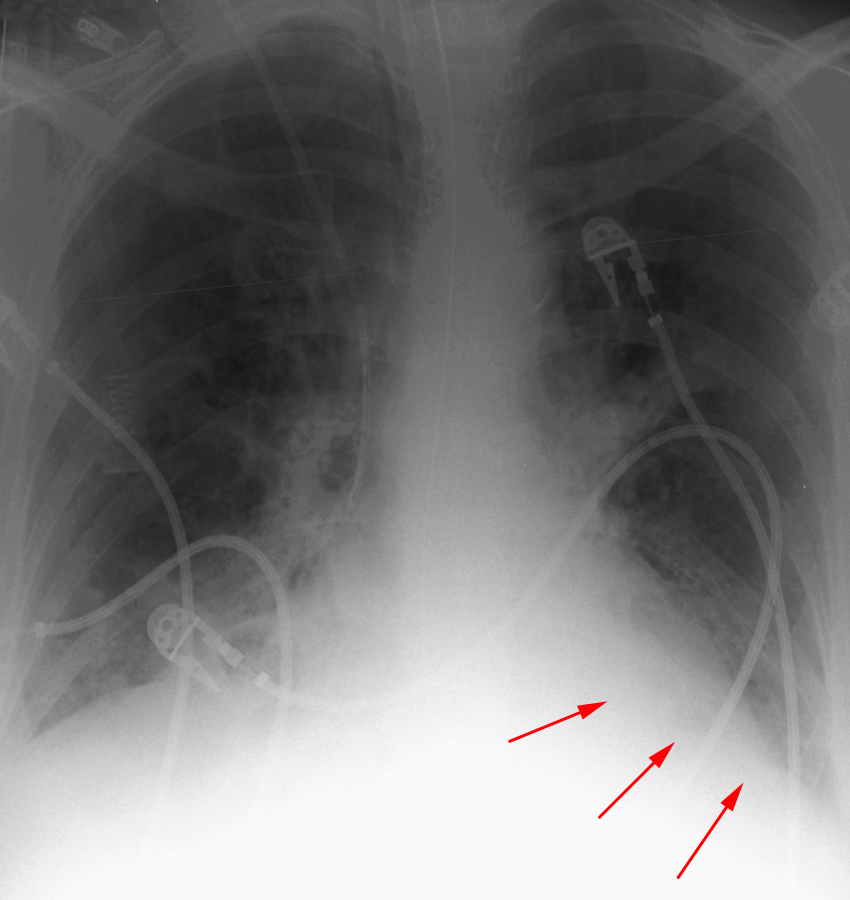
Radiographically, atelectasis may vary from complete lung collapse to relatively normal-appearing lungs. For example, acute mucus plugging may cause only a slight diffuse reduction in lobar or lung volume without visible opacity. Nevertheless, the physiologic effects can be significant. In the so called mucus plugging syndrome, the association of sudden hypoxia with a normal or quasi-normal chest radiograph can lead to the suspicion of a pulmonary embolus. Mild atelectasis usually takes the form of minimal basilar shadowing or linear streaks (subsegmental or "discoid" atelectasis) and may not be physiologically significant. Atelectasis may also appear similar to pulmonary consolidation (dense opacification of all or a portion of a lung due to filling of air spaces by abnormal material), making it difficult to distinguish from pneumonia or other causes of consolidation. The distinction between atelectasis and other causes of consolidation is important, and certain clues exist to aid in making that determination. Atelectasis will often respond to increased ventilation, while pneumonia, for example, will not. Crowding of vessels, shifting of structures such as interlobar fissures towards areas of lung volume loss and elevation of the hemidiaphragm suggests atelectasis. Another key for distinguishing between atelectasis and consolidation is recognition of the typical patterns that each pulmonary lobe follows when collapsing.

Left lower lobe atelectasis with lost of the hemidiaphragmatic shadow
(arrows).