ICU Chest Films > Lines and Tubes > Thoracostomy Tubes > Incorrectly Positioned Thoracostomy Tubes
Incorrectly Positioned Thoracostomy Tubes
![]()
Following the insertion of a thoracostomy tube, a chest x-ray should be obtained to verify its position. Tubes placed within fissures often cease to function when the lung surfaces become apposed (See x-ray below). Also, incorrectly placed tubes for empyemas may delay drainage and result in loculation of the purulent fluid. Determining whether a tube is anterior or posterior is often difficult with a single AP chest x-ray.
What
other view may help you determine if a chest tube is posterior or anterior?
PA
Lateral
Lordotic
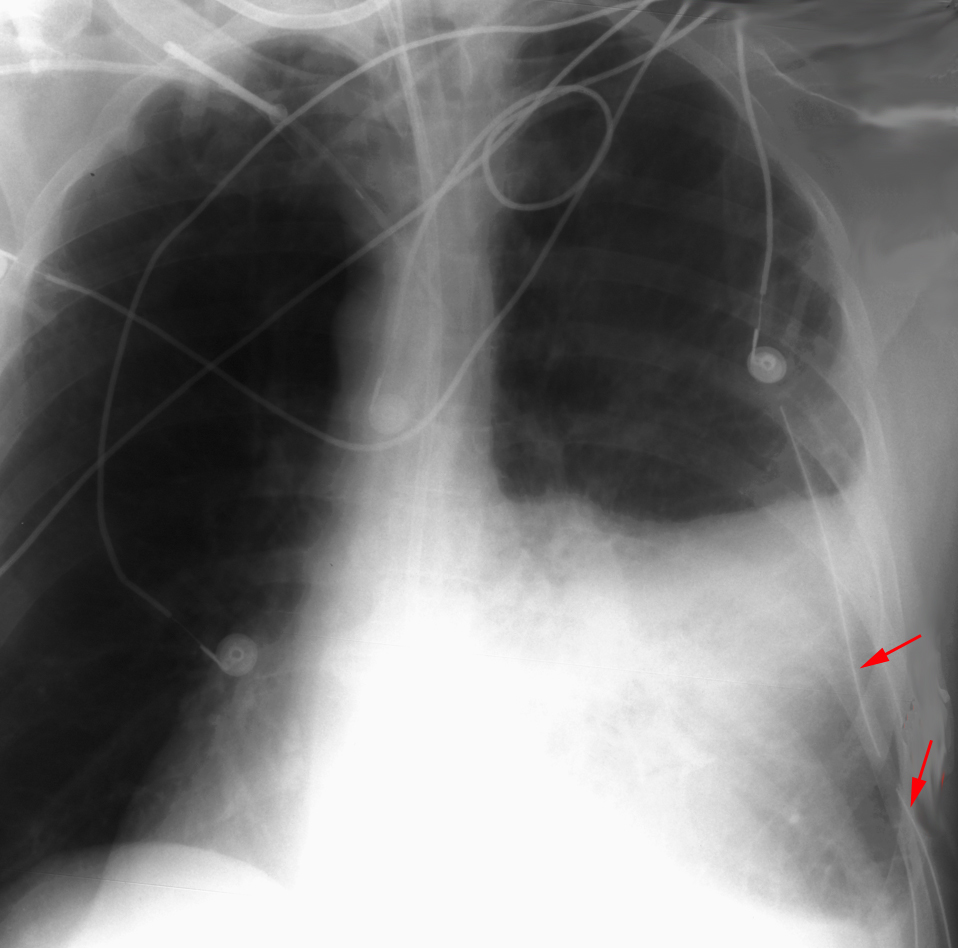
In order for thoracostomy tubes to function properly all of the fenestrations in the tube must be within the thoracic cavity. The last side-hole in a thoracostomy tube is indicated by a gap in the radiopaque line. If this interruption in the radiopaque line is not within the thoracic cavity or there is evidence of subcutaneous air, then the tube may not have been completely inserted.

This chest tube failed to remove