ICU Chest Films > Air in the Chest > Pneumomediastinum > Radiographic Appearance of Pneumomediastinum
Radiographic Appearance of Pneumomediastinum
![]()
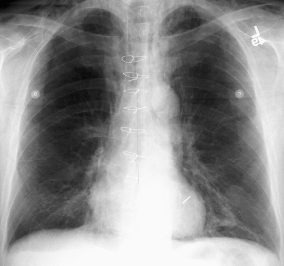
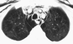
The radiographic appearance of pneumomediastinum results from air outlining structures which are not normally visible on chest x-ray. Pathognomonic signs of pneumomediastinum include lucencies around the great vessels, the medial border of the superior vena cava, and the azygos vein. Air may also be seen outlining the aortic knob, descending aorta, or the pulmonary arteries.


Notice the lucencies around the great vessels and superior vena cava seen
on both AP chest film (left) and CT (right).
Patients with posteromedial pneumomediastinum (usually due to esophageal rupture) may have dissecting air at the paraspinal costophrenic angle and beneath the parietal pleura of the left diaphragm. This is called the V-sign of Naclerio.