ICU Chest Films > Air in the Chest > Pneumothorax > Pneumothorax in the Erect Patient
Pneumothorax in the Erect Patient
![]()
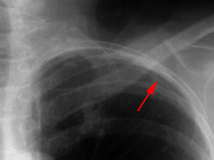
In the erect patient, air will rise to the apicolateral surfaces of the lung. An apicolateral pneumothorax appears as a thin, white pleural line with no lung markings beyond. The presence of lung markings beyond this line, though, does not exclude pneumothorax. This is especially true in the patient with parenchymal disease which may alter the compliance of affected lobes, making their collapse more difficult to detect radiographically. Parenchymal disease may also make visualization of the pleural line more difficult or impossible.

Apical pneumothorax, notice the absence of lung markings beyond the pleural
line.
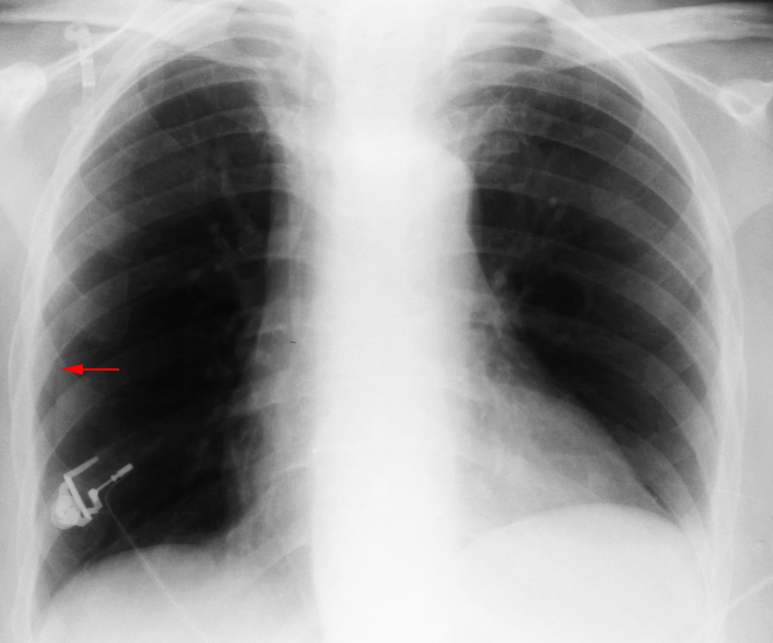
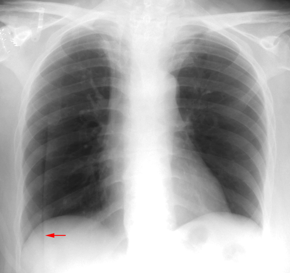
Skin folds on a patient can mimic a pleural edge and a pneumothorax. One can sometimes differentiate the two by noting that the skin fold line continues outside of the chest.


On the initial chest radiograph (left) the patient appeared to have pneumothorax.
Repeat chest radiograph (right) revealed that what has appeared as a pleural
line was a skin fold.